
by Heather Owen, DVM, MAV, CCRP, MT, CCFT, and Lisa Mason, DVM, CCRT, CVA, CVPP
Worried about recent reports on adverse events from anti-NGF monoclonal antibodies (mAbs)? Wondering when to pivot or pair with other therapies?
Dr. Heather Owen and Dr. Lisa Mason answer these important questions and more with the latest available information.
This webinar covers:
- How anti-NGF mAbs work
- What the latest literature reveals
- Reported side effects, including what to watch for in your practice
- Tips for monitoring osteoarthritis (OA) patients on mAbs
- Video tools for pain & joint assessment in OA
- Ideal patient profiles for mAb therapy
- Real-world cases to apply what you learn
Learning Objectives
- How anti-NGF mAbs reduce OA pain and where they may not be ideal choices
- Red flags for adverse reactions and tools for regular monitoring
- Step-by-step OA pain & mobility assessments (with video links)
- When to introduce photobiomodulation or PRP for safer, synergistic care
- Insights for cases you’re seeing today
Click here to download the slides and notes
Videos Referenced in the Webinar
×
×
×
×
×

×
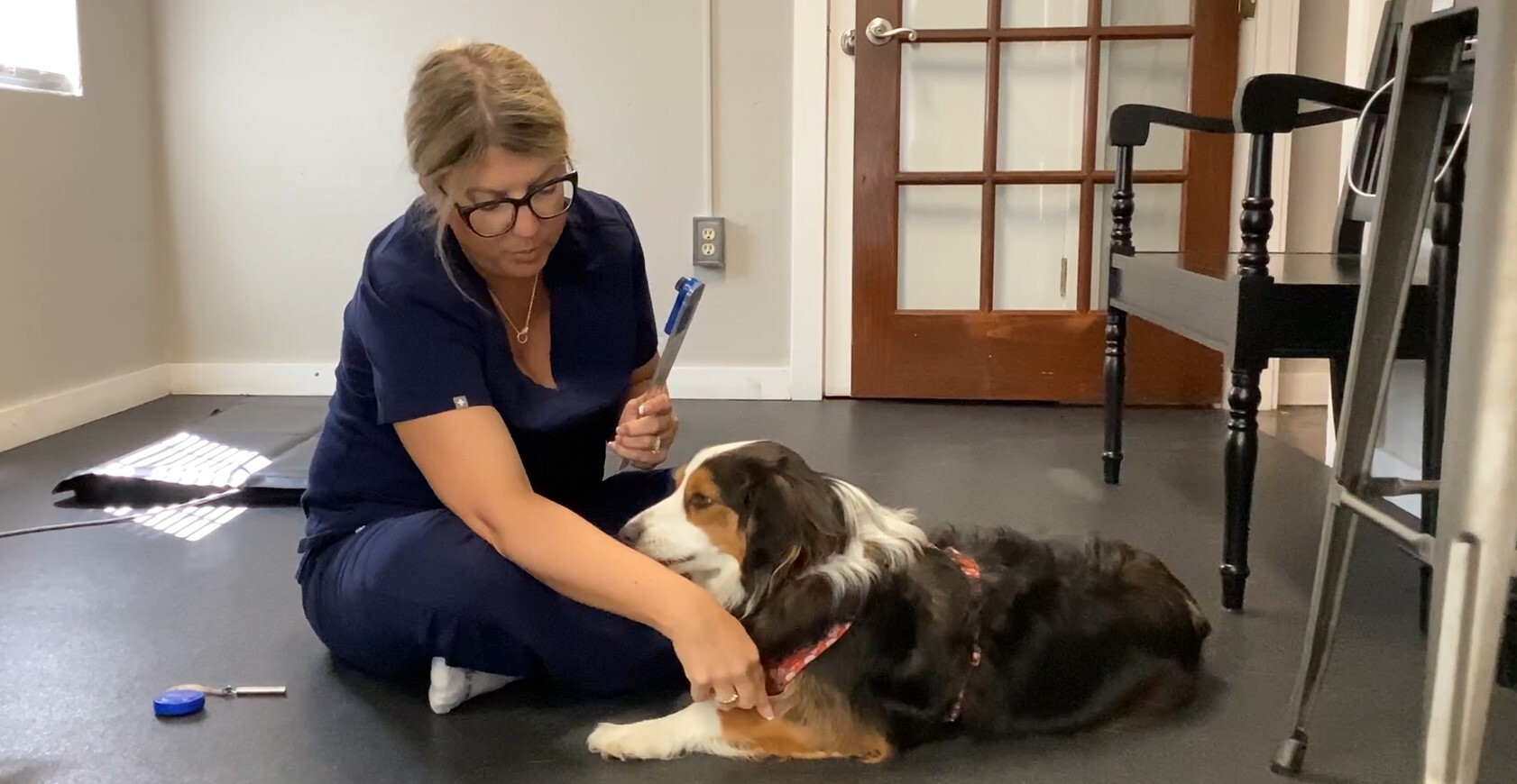
- 1. Goniometry and Thigh Circumference: The video clip demonstrates how to quickly perform goniometry (measuring joint angles in flexion and extension) as well as measuring the thigh circumference to provide objective measures to monitor patient progress or decline. This is a quick and objective test that can be done in your practice with minimal investment.
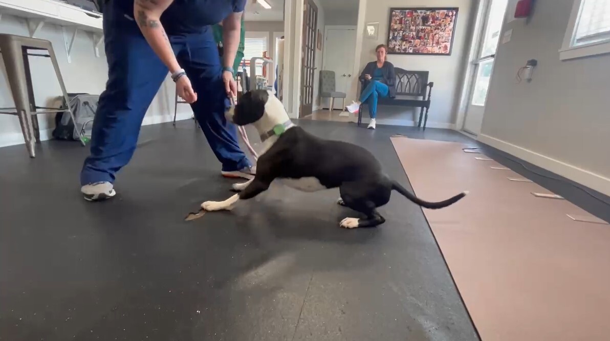
2. Transition Down to Stand and Sit to Down: Shows a patient performing Down to stand transition evaluation. In this video this patient struggles with full hip and stifle flexion and keeps her hips off of the ground. This is characteristic with hip arthritis. (PennHip confirmed moderate hip dysplasia in this patient). You can watch the patient push to a stand with front limbs to avoid hip extension.
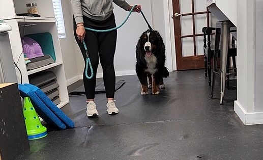
3. Lameness Testing 2: Shows a patient being videoed at a trot to view their lameness. In this video you can see elbow abduction with minimal elbow flexion. You can also appreciate swelling over both tarsal joints. This patient has confirmed elbow osteoarthritis.
4. Lameness Testing: Shows a patient with severe multiple joint disease with weakness. In this video we can see weakness but we also note the head bob classic with equine lameness (down on the sound), we can see minimal carpal flexion and extension, minimal elbow flexion and the view of the hind limbs demonstrates stifle and hip concerns as well. The stifles are collapsing medially, hips are in flexion with the pelvis tucked and the thoracolumbar spine is in flexion. This patient has confirmed elbow osteoarthritis, hip osteoarthritis, stifle osteoarthritis secondary to bilateral cranial cruciate ligament ruptures.
5. Neurologic Gait: Shows a patient with proprioceptive ataxia, which is a neurologic gait abnormality. There is hind limb ataxia, proproprioception deficits and hind limb paresis resulting in collapse.
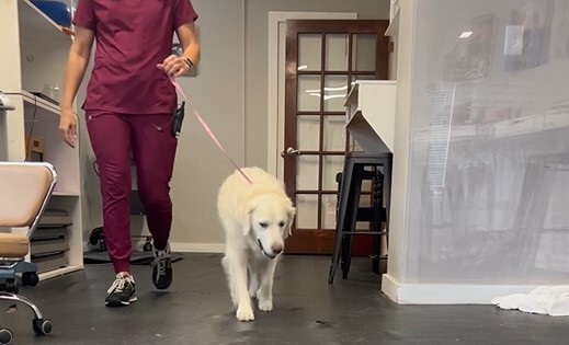
6. Transition Evaluation: A patient demonstrating how to observe a patient's transitions, from sit to down to stand on a carpet or non-slip floor. (also transition eval presentation). This patient demonstrates the classic front limb slide to down when elbow flexion is painful. This patient has confirmed early elbow osteoarthritis
Please note that the original speaker will not be available to answer questions submitted for on-demand webinars. Our medical team will answer any questions to the best of their availability, but this may not be possible depending on the question submitted.